Liver Conditions
Certain factors make it is very difficult to diagnose liver disease in dogs . * Symptoms of liver disease in dogs are difficult to pin point as they are subtle and vague and often mimic those relating to other diseases. * Liver cells can continue to perform their dedicated functions despite the liver mass being affected. * The liver has a great reserve capacity. * The liver can be affected by other diseases as it supports and is supported by many other organs and systems in the body. All these factors can lead to frustration for a veterinarian to diagnose whether the liver is affected and to what extent. One of the tools that the specialist will use is to get a complete chemistry profile of a blood sample. Although, clinical pathological tests and enzymology play a crucial role in arriving at correct diagnoses, it seldom indicates any deviation from a healthy condition of the liver. Enzymology is a branch of biochemistry that deals with the chemical nature and biological activity of enzymes. Another factor that complicates diagnosis of liver disease is that the levels of even enzymes that are specific to liver can be disturbed by secondary hepatic disease also. ALT (alanine aminotransferase) or SGPT (serum glutamic pyruvic transaminase) is a liver specific enzyme. It is concentrated in the cytosol and is released when localized liver cells die from infection or the interruption of blood supply. Serum levels increase two to three days after the liver has been affected and return to normal after a couple of weeks of treatment. Generally, two to three times the normal level is considered as insignificant and only a persistent increase is considered to be abnormal. ALT levels may go up to four to five time the normal level even in non-hepatic disorders like inflammation of the gastrointestinal tract, hemolytic anemia and heart failure. A dog undergoing treatment with anticonvulsants and glucosteroids or those that have an inhibited flow of bile may also show a moderate increase in ALT levels. AST (Aspartate aminotransferase) or SGOT (serum glutamic oxaloacetic transaminase) is another liver enzyme, also found in muscle tissue and red blood cells. An increase in levels of AST indicates a more severe liver disease than ALT. SAP (Serum alkaline phosphatase) levels increase in certain forms of cancers including liver cancer. Elevated levels of SAP are more significant in cases of feline liver disease than in dogs. GGT (Gamma-glutamyl transpeptidase) levels signify liver disease caused by blockage of bile ducts. Vague symptoms of liver disease in dogs like diarrhea, vomiting and anorexia are often misread as relating to indigestion and other mild ailments. Symptoms like jaundice, which are specific usually surface at a later stage of liver disease. In such a situation, despite the confusion over reading the results of laboratory tests and the accompanying emzymology, a complete chemistry profile is of utmost necessity to diagnose liver disease in dogs.
For more in-depth reading regarding Liver irregularities… please read the following sections below:
Protein is Essential for a Canine Liver Disease Diet
Canine liver disease diet should contain normal amounts of high quality protein, at least 20% of daily calories. The exception to this is if your dog has hepatic encephalopathy, a condition in which the liver disease has advanced so far that the brain has become affected. In this case, a low-protein diet is recommended. High-quality proteins are better digested and have an amino acid content close to the levels your dog needs. Foods that come from animals or from plants such as soy isolates, wheat gluten and dairy products are better tolerated than meat proteins in people which may be the case with dogs. Most veterinarians recommend that owners feed their dogs a mix of animal based and plant proteins since the use of soybean or lactose-containing dairy protein diets are not liked by some dogs and can cause diarrhea.
Non-Protein Calories
Non-protein calories help to prevent the use of protein (amino acids) for energy and reduce the need for your dog’s body to manufacture glucose in the liver by converting protein molecules (called gluconeogenesis). Normally, energy is from fat since it is a something dog’s like to eat and is a concentrated source of energy. Dogs with liver disease can tolerate larger quantities of fat in their diet (30 – 50% of calories).
Fiber and Canine Liver Disease Diet
Moderate amounts of soluble and insoluble dietary fiber can help a dog with liver disease. Soluble fiber such as beet pulp and gums lowers the production and absorption of ammonia and helps the growth of beneficial bacteria. Fiber (both soluble and insoluble) also helps your dog rid itself of bile acids. Insoluble fibers (lignin, cellulose, hemicellulose) help to normalizing transit time for feces, prevent constipation and bind toxins.
Vitamin and Herbal Supplements for a Canine Liver Disease Diet
A canine liver disease diet should include vitamin supplements that act as antioxidants. Liver diseases cause greater generation of free radicals and oxidant stress. Supplementation with antioxidants helps to reduce liver injury. B Vitamins: are often recommended at double the normal maintenance dose since this is a clinically supported approach in humans. C Vitamins: Vitamin C is a antioxidant and should be part of a dog’s diet with liver disease. Most dog foods meet the daily requirement for vitamin C. Do not overdose vitamin c since it could increase the intake of copper. Additional supplementation should only be necessary if your dog’s liver is having trouble with in case where fat is not be digested normally (fat mal-absorption). Vitamin E: may prevent canine liver disease from getting worse by reducing free radical or oxidant injury. A water-soluble form of Vitamin E is preferred, since the absorption of fat-soluble vitamins may be decreased in some forms of liver disease. Vitamin K: helps with blood clotting and is recommended in cases of chronic liver disease. The liver produces clotting factors, and it does not produce or store vitamin K as well when it is diseased. Zinc: A canine liver disease diet should also be supplemented with zinc since it is an anti-oxidant. It also reduces the risk of abnormal brain function caused by passage of toxic substances from the liver to the blood (called hepatic encephalopathy). Zinc reduces the accumulation of copper in the liver. Adenosylmethionine (SAMe): may be helpful in reducing liver injury. Normally produced by the liver, SAMe is necessary for many functions of liver cells. It is also an anti-oxidant. Providing your dog with an oral supplement helps to to improve antioxidant function and has anti-inflammatory properties. Phosphatidylcholine (PC): is a components of bile required for normal bile acid transport and a building block for cell membranes. There are no clinical studies showing that it is helpful in dogs (it is helpful in humans). Silymarin: a component of milk thistle is thought to have antioxidant and helps with free radicals for various types of liver disease. It also helps with toxicity. There are a few studies that support its use for dogs. Suggested doses range from 50 to 250 mg/day A supplement such as Pet Alive Liver-Aid formula combines many of these recommended supplements and is worth exploring. Check with your veterinarian so that they can track your progress.
Minerals and Supplements to Avoid
Potassium: Diets for dogs with liver disease should avoid potassium. If your dog is not eating and therefore not getting any potassium your veterinarian may choose to give your pet the mineral through intravenous fluids. Sodium: Moderate restriction of dietary sodium if recommended for dogs that have a lower than normal appetite or if your pet has hypertension. Copper: Diets low in copper are recommended for certain breeds such as Bedlington Terriers, Doberman Pinscher, Skye Terrier and West Highland White Terriers, Airedale Terrier, Bobtail, Boxer, Bull Terrier, Bulldog, Cocker Spaniel, Collie, Dachshund, Dalmatian, German Shepherd, Golden Retriever, Keeshond, Kerry Blue Terrier, Pekingese, Poodle, Samoyed, Schnauzer, Wirehaired Fox Terrier. These breeds are prone to a form of liver disease called copper storage disease in which too much copper accumulates in the liver and causes problems with liver function. It should be noted that dogs with copper storage disease should not be given Vitamin C because it may increase the damage to the liver. If you have these breeds your dog’s diet should center around foods low in copper such as beef, cheese, eggs and tomatoes. Avoid lamb, pork and duck. Check with veterinary nutrition experts for a complete list.
Canine Liver Disease Diet Prepared at Home
These two diets were created by veterinary professionals for dog’s with liver disease problems:
| Home Canine Liver Disease Diet #1 | (1000 g diet) |
|---|---|
| Chicken, breast with skin | 220 g |
| Rice, Cooked | 680 g |
| Carrots (boiled, drained) | 60 g |
| Wheat bran | 20 g |
| Rapeseed oil | 20 g |
| Home Canine Liver Disease Diet #2: | (1000 g diet) |
|---|---|
| Beef, minced meat, 15% fat | 100 g |
| Tofu | 400 g |
| Rice, cooked | 440 g |
| Carrots (boiled, drained) | 30 g |
| Rapeseed oil | 20 g |
| Wheat bran | 10 g |
In addition to either of these diets give your dog a supplement that contains the ingredients indicated above. Make sure that any selected supplement is low in sodium and copper. A good choice made specifically for this purpose is PetAlive. Check with your veterinarian to see if it would be helpful for your dog’s specific condition.
Commercial Canine Liver Disease Diet
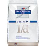 Canine liver disease diet formulated by Hill’s Prescription Diet l/d. This and those made by other pet food manufacturers use higher quality protein to reduce the amount of work done by the liver and to aid in the regeneration of liver tissuye. They contain antioxidants such as Vitamin C and Vitamin E to avoid oxidative stress.
Appetite Problems
If your dog has little or no appetite you can try warming your dog’s food a bit to make it more appetizing. Try feeding small amounts several times a day. If that doesn’t work, your dog may need to be force fed with a syringe or a feeding tube may need to be placed. Your dog also needs to be getting plenty of fluids. If your dog is not drinking on his own, he may need to get IV or subcutaneous fluids to prevent dehydration.” Another excellent link on how to overcome feeding issues (no appetite) with Liver Disease http://www.scamperingpaws.com/liver-disease-dogs/hope-for-healing-canine-liver-disease.shtml This is some of what is included in the link above:
- Try warming up the food to room temperature, or slightly warmer. Heating up food can help enhance its scent. This may be enough to persuade your dog to at least try the food.
- Try serving the food cold. If your dog is feeling nauseous, strong scents may turn him off completely. So try serving chilled food instead. Offer a small bite-sized morsel first to try and entice him to eat. My dog accepted cold food far more often than warmed food.
- Bake canned dog food. Invert a can of dog food until the food comes out in one piece. Then slice the food as thin as you can and place them on a cookie sheet. Bake at low temperatures until the slices are completely dry.
I had good luck using an egg slicer to quickly slice up firm, canned food (just cut to fit, then slice). If the canned food is softer, the egg slicer won’t work – at least it didn’t for me – and I found that a pastry cutter worked better (although it slices the food a little thicker than I would have liked).
- Cook – and grocery shop. I tried scrambled eggs (sometimes with ham or cheese), fried eggs, hard-boiled egg yolks, ground chicken / turkey / beef simmered with rice; roast beef; roast chicken and flavored chicken wings from the grocery store; chicken pot pie; oatmeal; pretty much anything (note: many, many times my dog refused the food I had prepared, so it became my lunch or dinner).
- Offer very small quantities. When my dog was feeling nauseous, a larger amount of food made it worse. Sometimes he would eat if I offered a teensy scarce 1/2 teaspoon of food at a time (at times, he would eat 1/4 cup or more of food in this way!).
- Offer food throughout the day. It’s hard to know when your dog will be feeling inclined to eat… so if it’s possible, offer him food whenever you can. You never know when he’s going to accept.
- Be creative in how you offer food. Sometimes we offered it on a fork, on a spoon, or offered fingerfuls of food. My dog would occasionally eat kibble off the floor, if I didn’t put down more than 3 or 4 kibble at a time. And sometimes he’d eat if I put a small amount of food beside him (not in front of him), so that he could easily turn his head and eat it when he wanted.
- Don’t force him to eat — it might make him turn away in the future. If he’s not eating, he might still be feeling too nauseous to eat. You don’t want him to associate the food with feeling bad. Just back off and try again later.
This topic is as big as the states of Alaska and Texas combined. I really cannot do it justice in a short essay, but I think it deserves some mention, if only to give the pet owner some understanding as to the difficulties involved when a veterinarian is faced with a case of possible liver failure. If you asked ten people on the street what they knew about “liver”, I would bet that the only consistent answer you would get is that it tastes really bad unless the cook really knows his stuff. The best description of the liver I can give you is that this organ is the main industrial centre of the body. The liver processes raw materials, manufactures the building blocks of the body, recycles the old to make new, and detoxifies the industrial waste of the body. In short the liver is involved in just about every biochemical process required to run e body. As a result of this relationship, liver disease can affect just about any other part of the body and thus the symptoms of liver disease are typically unpredictable and non- specific. Furthermore, because the liver acts as a “biochemical cross roads” for the body, it is affected by a wide range of diseases, including viral and bacterial infections, degenerative and neoplastic disease, and toxic insults. It is estimated that three per cent of all disease seen by veterinarians is liver based. The liver has a double edged nature which, while being life preserving, makes diagnoses and treatment of liver disease extremely difficult. The liver has a tremendous reserve capacity, which means that it can easily perform it’s duties with up to 70 to 80 per cent of the liver mass affected by disease. While it certainly is a benefit that our liver can keep us alive despite an overwhelming infection or a massive tumour, it also means that the disease is well advanced and possibly untreatable before any symptoms are noted. We all know that disease is most easily conquered early, but the very nature of the liver makes this an impossible task. One thing about livers though: they are the only organ in the body which is capable of complete regeneration and thus is we do manage to successfully treat the disease, there is a chance of complete recovery. Because of the complexity of this topic, I am going to cover it using very abbreviated point form. I will try to skip over the experimental theories and the more esoteric points and keep to the meat of the topic. Common Presenting Symptoms All, some, or only one of these signs may be present. Intermittent recurrent abdominal or gastrointestinal upsets. loss of appetite, vomiting, diarrhea, constipation. Progressive depression or lethargy. does not want to play anymore or refuses to go for walks. Swollen belly with a “fluid filled” look. This is also known as ascites and is actually fluid accumulation in the belly due to circulation alterations in the abdomen. Pale gray feces. Bile pigments are what gives poop it’s characteristic brown colour and if the liver is not processing bile properly, the feces will not get their colour. Orange urine. The improper processing of bile results in the excretion of bilirubin in the urine in high amounts, thus orange urine. Jaundice, also known as icterus. Any pale or white skin or visible tissue takes on a yellow hue. Again the biliary pigments are accumulating in the body because the liver is not processing them. Rarely: bleeding problems. Many of the proteins required for proper blood clotting are created in the liver. Remove these proteins and blood clotting decreases. Hepatic encephalopathy, or severe neurological signs. behavioural changes, seizures, aimless pacing or circling, head pressing. May be associated with meal time. Pain associated with the abdomen. This is due to the stretching of the liver capsule. May be noted when the dog is lifted around the belly or when the veterinarian probes (palpates) the abdomen. The veterinarian may also notice a swollen liver while palpating with some of the more acute liver diseases. Chronic weight loss or wasting. The liver processes all the building blocks. If it fails to process, the body fails to maintain itself. Increased water consumption and urination. Most likely due to dramatic shifts in serum and kidney salt balances. May be behavioural too. Points on notable symptoms. 1. Bile pigment processing. Bile is a complex mixture of organic and inorganic compounds. It is primarily responsible for alkalizing the intestinal contents (acidic from the stomach),emulsifying the dietary fat, and prevention of putrefaction of digestive material. Bilirubin, one of the bile pigments, is derived from the break down of hemoglobin, the oxygen carrying molecule carried in our red blood cell. Bilirubin is quite toxic, but it usually binds to a protein called albumin, which harmlessly carries it to the liver for detoxification and excretion. Albumin is made in the liver. Liver failure results in poor bilirubin processing and decreased albumin manufacturing, which results in a dangerously high level of free floating bilirubin. The liver excretes the bilirubin after binding it to an amino acid into the bile duct system. Eventually the conjugated bilirubin enters the digestive tract, where the intestinal bacteria break it down to a harmless product called urobilinogen. Urobilinogen, after complete digestion in the intestines, is brown, therefore the feces tend to be brown. Jaundice, also known as icterus, results from the accumulation of conjugated and unconjugated bilirubin in the body tissues. This becomes visible to the veterinarian, especially around the whites of the eyes and on the pale areas of the gums. 2. Important biological functions: • a. hormone metabolism. The liver is both the target organ for many of the body’s hormones and the recycling centre for most of the hormones. Some of the symptoms stemming from liver failure may mimic a major hormonal imbalance. • b. vitamin metabolism. Practically all the vitamins consumed in our diets are either directly involved in liver function or require liver aided transformation to be used in the body. This includes Vitamin C, the B vitamins, Vitamins A, D, E and K. Vitamin K is important to maintain blood clotting and requires hepatic transformation from the inactive form to the active form. • c. Red blood cell maintenance. In the mature dog the liver plays an active role in the removal of aged or damaged red blood cells from circulation. It is also active in the metabolism of hemoglobin and the storage of iron. Abnormalities in red blood cell structure is one of the harbingers of liver disease. Anemia may be present in chronic liver disease. • d. Hemostasis or blood clotting ability. Most of the proteins involved in the creation of a functional blood clot are made in the liver. The clotting system is an extremely complex, interlocking system ; remove some of the factors involved and you end up with a tendency to bleed or hemophilia. • e. Carbohydrate and fat metabolism: Sugars, or carbohydrates are the basic fuel of the body. The liver is the primary centre for processing of the sugars into the form immediately required. The liver is also responsible for the destruction of insulin, the hormone directly involved with the cellular absorption of blood sugars. Alterations in liver function often do not affect blood sugar levels until much of the liver has been destroyed. Fat metabolism is extremely complex due to the vast number of functions fat carries out in the body. The liver sits at the centre of those many functions. Cholesterol is probably the most common fat based product in the body, being the major component in the cell wall, the basis for the steroid hormones and bile pigments, and the precursor of vitamin D. Any disease in fat metabolism can adversely affect the liver, and any disease in the liver can result in problems in fat metabolism. An example of this is the “fatty liver syndrome” we see in cats, whereas the rapid mobilization of fat stores during starvation results in an overtaxed liver and eventually liver failure. Protein synthesis: The liver manufactures many of the proteins involved in the body functions. The major protein is albumin, which is required for transport of many nutrients and toxins (i.e. bilirubin). Albumin is also responsible for keeping the serum concentration constant, which is important with regards to serum fluid and salt balance. (also known as “oncotic pressure”) Also synthesized in the liver is the globulin series, which are responsible for numerous biochemical reactions throughout the body. Elevations of select globulins may indicate a particular hepatic pathology. The building blocks of proteins are the amino acids. The liver is also primarily involved in processing of dietary amino acids to modify them into required or useful forms. Some of the amino acids require direct hepatic metabolism, while others can be used by the body unchanged. Experimental efforts have been made to diagnose and track liver disease based on the relative proportion of the various amino acids to each other. In liver failure the amino acids requiring hepatic alterations prior to use should climb in concentration as compared to those amino acids unaffected by the liver. Important Liver Enzymes Traditionally the medical practitioner has measured the relative concentration of several enzymes which may indicate alterations in liver health. The following enzymes typically change values in the face of liver failure: Alanine Aminotransferase: ALT. Liver specific in the dog and cat. Cell damage will cause elevations of A-LT due to leakage. The elevation of the enzyme correlates with the number of cells damaged. Falling levels of ALT may indicate recovery or may indicate a failing number of functional liver cells. Rapid increases in ALT may indicate an acute process, while slow increases may indicate bile duct obstruction. Aspartate Aminotransferase: AST, an enzyme seen in the liver, heart, kidney, skeletal muscle and brain. The half life of the AST in the blood stream is much shorter than that of ALT, therefore the values of AST tend to drop more rapidly once liver function is resumed. AST elevations and ALT elevations should parallel each other in liver disease. Alkaline Phosphatase. This enzyme is present in many tissues, therefore it is not very specific in liver disease, but it appears very early in the progress of liver disease, therefore it is considered quite sensitive. ALP tends to be slightly more specific in the cat, but not quite as sensitive. A similar enzyme or isoenzyme is secreted as a result of high levels of cortisone, therefore an effort must be made to separate Cortisole induced ALP or CALP and normal ALP. Liver ALP is released from the liver when many anticonvulsant drugs are administered to the dog. A similar sensitivity has not been noted in the cat.. This must be taken into account when evaluating ALP levels. ALP levels typically are greatly elevated in the young, growing animal and therefore a veterinarian should not mistake any elevations as disease in a young animal. Gamma Glutamyltransferase GGT: This enzyme is has it’s highest concentration in the kidneys and pancreas, but it is also found in the liver and other organs. The major proportion of GGT in the serum seems to come from the liver. Elevations of GGT in disease seem to stem from new synthesis rather than leakage, therefore the changes seen due to disease are not spectacular. Large elevations of GGT are more commonly associated with pancreatitis and bile duct obstruction. Bile Acids: These series of organic acids circulate almost entirely in the localized blood flow between the intestines and the liver (a.k.a.: the Portal system). The flow is typically from the liver, into the bile duct system, then excretion into the intestines to aid digestion after a meal, to be re- absorbed into the portal system and recycled by the liver. Very little of the bile acids escape form the portal circulation system into the rest of the body. Leakage is considered abnormal and is a sure sign of a liver abnormality. This is one of the most sensitive tests available to diagnose liver disease. While the liver does actually manufacture this product, it has tremendous reserve capacity and can easily meet the bodies demand for bile acids despite severe disease. As a result of this reserve, the bile acid levels do not typically drop due to liver disease. Ammonia and Urea: Ammonia is a by product of digestion of protein in food and the catabolism of nitrogen based organic materials in the body. Eighty per cent of ammonia is delivered to the liver and converted to urea. In patients with liver insufficiency the ammonia is not detoxified to urea, but enters the circulation to act as a central nervous system depressant. In patients with a severely reduced liver function we may see a true intolerance of ammonia and thus neurological signs after a heavy protein meal or we may see substantially reduced urea levels. This is a late sign in liver disease, only seen after 60 to 70 per cent of the liver function is gone. Ascites development: This is the accumulation of fluid in the abdominal cavity and results from several factors. Simply put liver disease tends to alter the blood pressure in the portal system, the albumin and salt concentration in the serum, the water retention in the body, the function of the surrounding organs and the permeability of the portal vessels. As a result of all these factors, fluid tends to build up in the abdomen and the animal gets a big, swollen, fluid filled belly. Electrolyte and Acid-Base disorders. Common side effect of liver disease due to a multiple of factors leading from metabolic disorders. Enough said. Gastrointestinal Ulceration and Hemorrhage: Again a sequellae to liver disease which may confuse the veterinarian. He may think he is treating a simple ulcer and miss the liver disease. Hepatic encephalopathy. Simply described as severe neurological dysfunction due to advanced liver disease. Has been linked to the accumulation of biological toxins, including ammonia, alterations in the blood brain barrier, alterations in the neuroreceptors in the brain, and decreased blood sugar. Diagnosis of Liver Disease Having gone into such detail prior to this, I hope this section will be short and to the point. I hope most of my statements here will naturally flow from points made above. 1. Examination, specifically noting signs which may indicate liver disease. Periodic ascites, intolerance of a high protein diet, icterus, chronic weight loss, abnormally coloured feces or urine, bleeding disorders, chronic illness, and all that has been mentioned above. Sometimes urinary crystals formed from the improperly metabolised proteins and amino acids may indicate liver disease. 2. Extensive blood work:
-
- A complete blood count to check for anemia and blood cell abnormalities.
>
- A complete chemistry screen, including ALT, ALP, AST, bilirubin, glucose, urea, electrolyte levels, albumin, globulin and bile acid levels. The bile acid levels should be checked on a empty stomach and two hours after feeding. All these values , with the exception of the bile acids, usually are included on a standard Small Animal Data Base Screen.
- A complete urine analysis. Check urobilinogen levels, bilirubin levels, glucose levels, protein levels. Again all this is usually on a standard urinalysis panel.
- Radiograph the abdomen. X rays can show increased liver size, decreased liver size liver abscesses, abnormal mineralization , and circulatory abnormalities (using special dyes)
- Ultrasound the liver. Perfect technique for visualizing the circulation of the liver, the bile duct system, the density of the liver tissue, the size of the liver.
- Biopsy of the liver. While this is a surgical technique, it is the ultimate for diagnoses, since it allows us to directly examine and test liver tissue, give an absolute diagnoses and hopefully a final treatment regime. Biopsies can be taken by full laparotomy, where the surgeon actually looks at the liver and removes a small piece, or they can be done by a biopsy needle guided by ultrasound through the body wall. The liver will regenerate any piece removed, therefore liver biopsy is usually a low risk procedure in capable hands.
Specific Diseases of the Liver Infectious Hepatitis Typically caused by either an adenovirus or a herpes virus. Transferred from dog to dog by oral contact and ingestion. Usually only causes a transient non specific illness characterised by lethargy vomiting, diarrhea and fever. Sometimes develops into a full blown case of severe hepatitis with many of the symptoms previously noted. Treatment is geared to support while the body fights off the bug. Prevention is by vaccination. Another syndrome has been seen in England called Canine Acidophil hepatitis. Typical signs of hepatitis are present, but the case may take a very chronic course, lasting over a period of years. No specific viral organism has been identified, therefore no vaccine or treatment is available. No specific virus causes hepatitis in cats, but the feline corona virus responsible for Feline Infectious Peritonitis will cause a hepatitis in some cases. Diagnoses by biopsy. No treatment. Mediocre vaccine. Several bacterial causes of hepatitis are known. Treatment is based on a proper diagnoses and appropriate antibiotic use. There is good proof that the bacteria is a normal inhabitant of the liver and only becomes a problem when the liver is injured form other causes. There are notable exceptions. Blastomycosis, histoplasmosis and coccidiomycosis are fungal infections seen in various parts of the country (usually associated with river systems) Difficult to treat. Leptospirosis is a bacterial infection common in wildlife and transferable to domestic animals and people through contaminated water. Dangerous, possible fatal, but the vaccine is quite good for prevention. Tuberculosis is still around and is considered transmissible to humans. Certain parasites will infect the liver. Typically the likelihood of parasitic infestation depends on the area you live in. Diagnoses is often based on symptoms, fecal examination, and standard diagnostic techniques for liver disease. Treatment is the use of appropriate parasiticides. Liver Disease Secondary to other Disease Acute pancreatitis: the close proximity of the pancreas to the liver and the bile ducts results in some degree of hepatitis whenever there is a case of pancreatic inflammation. Treat the pancreatitis and the liver disease will regress. c bowel disease: the chronic inflammation of the bowel allows portal absorption of toxic intestinal products and bacteria. treat the colitis. Shock, anemia, and congestive heart failure. All these result in severe loss of blood circulation to the liver and lack of oxygen. The liver disease is rarely of primary concern as the primary causes of the problem are most likely going to kill the animal prior to liver failure. Abdominal trauma: tears, bruising, biliary leakage, hepatic bleeding. Correction of these problems would require surgical intervention, assuming a timely diagnoses. Simple bruising of the liver will heal unaided, with only a transient increase in the hepatic enzymes. Feline Hyperthyroidism: many of the symptoms of hyperthyroidism and hepatitis are the same and in fact the hyperthyroidism will cause elevations in the liver enzymes. The thyroid level of any cat presenting with symptoms suggestive of liver failure should be checked. The hepatitis will resolve once the hyperthyroidism is treated. Chronic Hepatitis Copper storage diseases in Beddlington terriers, Doberman pinschers, and West Highland white terriers. These are all genetically inherited diseases which result in abnormal and toxic levels of copper to be stored in the liver. The course of the disease is variable, some presenting with acute hepatitis, many presenting in end stage cirrhosis of the liver. Diagnoses is based on liver biopsy. Treatment requires the use of copper binding drugs, anti inflammatory to decrease liver inflammation, dietary modification to limit copper uptake. Chronic Active Hepatitis: In humans there is a chronic form of hepatitis characterised by chronic elevation of liver enzymes and biopsy samples showing scarring and active inflammation. The underlying cause for this entity falls into one of three categories: viral induced, toxin induced, and immune mediated. There is some question as to whether a similar syndrome exists in dogs. There has been cases which did show chronic elevation of the liver enzymes over weeks to months), symptoms characteristic of liver disease ill defined malaise), and a response of anti inflammatory treatment to limit the ongoing inflammation and scarring of the liver. At this time recommendations for treatment are that moderate or intermittent disease should only receive supportive therapy or basic nursing, while deteriorating chronic cases should receive steroid based anti inflammatory. If the case shows poor response, biopsies should be referred to a pathologist for evaluation in an attempt to find the underlying cause. In some cases it may be necessary to use strong immune suppressant drugs to stop the destruction of the liver. Leptospirosis associated chronic hepatitis: An example of bacterial infection leading to chronic disease. Diagnoses by biopsy and identification of the pathogen. Treatment by antibiotics. Infectious Canine Hepatitis associated chronic hepatitis. Exposure to the virus responsible for ICH leads to chronic active hepatitis due to an ongoing immune system malfunction. Diagnoses by biopsy and the use of special stains to demonstrate the viral antigens in the liver. Lobular dissecting hepatitis: rare disease diagnoses by biopsy. Hepatoportalfibrosis: Disease primarily of the blood supply to the liver. Diagnosed by very specialized radiograph techniques which measure and visualize the blood flow through the liver. Biopsy critical for diagnosing location of lesion. Toxic liver injury: Primary disease is caused by the ingestion , injection, or inhalation of a toxic substance which adversely affects the liver. Due to the central nature of the liver with regards to detoxification of chemicals, it is no surprise that many are harmful to the liver. Factors contributing to the disease are: females more susceptible, fatty diets more dangerous, continuous exposure, high levels of exposure to toxins. Exposure results in death and inflammation of the liver cells, followed by replacement of damaged tissue by fibrous scarring. This can be a self perpetuating cycle, resulting in cirrhosis of the liver. Toxins include many common drugs, such as acetaminophen, ASA, anabolic steroids, chemotherapy drugs, some antibiotics, glucocorticoids, anaesthetics, parasite control drugs, and phenylbutazone. Some of the drug induced hepatitis is a predictable side effect of the drug, while other incidences of hepatitis are considered an unpredicted or abnormal side effect of the drug. This is difficult to diagnose unless there is a known exposure to the drug or toxin and the appropriate tests are taken. Biopsy will confirm liver destruction, inflammation, and fibrosis, but it will not single out the causative agent. Glucocorticoid Hepatopathy dogs seem abnormally sensitive to glucocorticoid drugs (“cortisone”) and will develop typical lesions in the liver after multiple dose therapy or long term over production of intrinsic cortisone by the adrenal gland (Cushing’s disease). Lesions are fairly typical and the rare animal which shows liver associated symptoms during glucocorticoid therapy will improve with the removal of the steroids. Liver associated lesions may take weeks to months to heal. Anticonvulsant associated hepatopathy; Phenobarbital, primidone, phentoin. May cause liver disease in 6 to 15 % of all dogs on anti-convulsant therapy. Inflammation seems related to dose. Degree of disease is variable and unpredictable. Diagnoses based on history, symptoms, laboratory tests, and biopsy. Treatment is removal of offending agent. Cirrhosis: This is the end point of chronic, active hepatitis. The cycle is one of liver cell death (due to insult, either toxic, viral, or immune mediated), followed by inflammation and scarification. The end [[[[ requires all previously noted techniques. No treatment is possible and ongoing palliative and dietary care is the only treatment option.. Noninflammatory Liver Diseases Portal vascular abnormalities Usually a portal-systemic shunt which allows blood to pass from the digestive tract directly into the general circulation without being detoxified by the liver first. Usually a congenital defect restricted to young dogs and puppies, but can be the result of hepatic cirrhosis. Symptoms are never consistent, but many dogs are young, malnourished, chronically sick, poorly tolerant of toxins, drugs, and anaesthetics, and tending to eat strange items (pica). Diagnoses is based on physical exam, history, laboratory tests, and specialized X rays showing blood flow through the liver. Treatment is surgical correction of the circulatory abnormality to force the blood into the liver prior to it entering the general circulation. Hepatic Lipidosis Most common form of severe liver disease in cats. Most often seen in obese cats suddenly subjected to dietary deprivation. May also be associated with diabetes mellitus, drug injury and toxicity. Thedisease seems to result from the sudden mobilisation of the bodies fat stores which quickly overwhelms the liver’s ability to process the raw fat into useful nutrients. The fat accumulates in the liver rapidly and causes acute liver failure. The end result is a swollen, greasy liver which is fragile and yellow to see. The cats present with complete lack of appetite and many signs of acute liver failure. Treatment is based on the provision of a highly nutritious diet to provide the energy required to run the body, stop the ongoing mobilisation of the fat stores, and drive the liver to decrease the fatty accumulation in the liver. Treatment is difficult and a long process. Hepatic Cancer (Neoplasm) Falls into two categories: primary or originally stemming from liver tissue or secondary; originating in some other part of the body and spreading to the liver through the circulation system. Primary liver cancers can stem from exposure to toxins (oncogenic compounds) which attack the liver full strength, since the liver is the primary detoxification centre of Secondary cancers may stem from any part of the body, but the liver is a favourite destination for metastatic cancer because of it’s central function in the body and the micro- capillary network which makes up the circulation passing through the liver. Primary liver cancer is usually quite advanced prior to diagnoses and tends to metastasize to the rest of the body very early in the course of the disease. Keep in mind the liver can function with less than one third of it’s volume still operating, therefore liver cancer can be very advanced before any symptoms are noted. Treatment is usually pointless, but would be based on diagnoses of the specific cancer and the use of appropriate chemotherapy agents. Basic Points for Treatment of Liver Disease
- Removal of toxic agents. Identify and remove any drug or toxin which may potentially hurt the liver.
- Rest and confinement. This will help divert body resources to the healing process at the liver and reduce discomfort caused by inflammation of the liver such as painful belly, nausea, malaise.
- Dietary management: Extremely important. The goal is to provide all the necessary nutrients which may be lost due to failure of liver processing without overtaxing the liver with regards to processing of dietary intake. High levels of top quality protein to provide the essential amino acids in an easily digestible carrier which will not produce high levels of ammonia during digestion. Cottage cheese is good, meat tends to produce high levels of ammonia. High level carbohydrates to drive the metabolism of the body, essential fatty acids not less than 6% of the daily intake, and a good mineral and vitamin supplement. Force feeding may be necessary.
- Control of ascites and water retention. Reduce sodium intake. Diuretics will help in resistant cases.
- Control concurrent infections with antibiotics.
- Deal with the concurrent medical problems as they crop up. Remember that the dog may develop bleeding problems, malabsorption problems, and neurological problems. Each separate problem has to be dealt with both individually and as a part of the whole disease entity. Neurological symptoms such as coma need to be addressed aggressively with a combination of therapies.
I realise that this is a long, possibly boring paper that is far from complete. I cannot emphasize too strongly how difficult this topic is. I gleaned this information from a single source Textbook of Veterinary Internal Medicine edited by Stephen J.Ettinger 1989. Liver Cleansing Diet For a diet that is useful for dogs with liver disease, those on extended medical treatment and dogs with epilepsy or seizures, the following link will provide a healthy home cooked diet. Please discuss this diet with your veterinarian before using it. It is a balanced meal designed by Dr. Jean Dodds and has been used by committee members for several years as a supplemental diet during illness. It is approved by The Epilepsy Foundation and others. http://www.canine-epilepsy-guardian-angels.com/liver_diet.htm